|

|
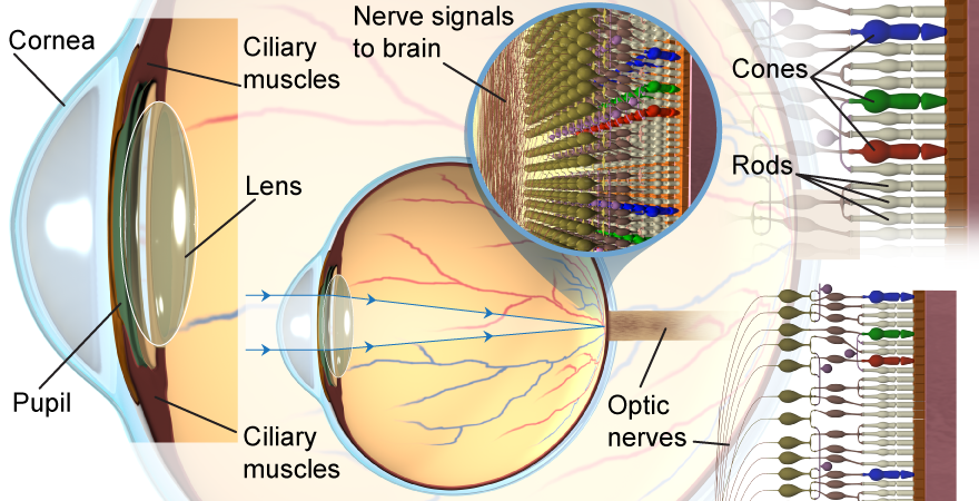
You probably take your eyes for granted. But how do they work? Your eye is very similar to a magnifying glass: A converging lens at the entrance to the eye focuses light into an image on the back of the eye (called the retina). 
 |
The eye has two different converging lenses that work together: the cornea and the lens itself. The cornea actually bends the light more than the lens, but it is the lens that can change its shape to focus the light from different distances onto the retina. 
|
How does your eye manage to see both in bright sunlight and dark nighttime? The pupil is an entrance hole that can vary its size to change the amount of light entering the eye. The iris dilates—opens the pupil wider—in dim conditions to let in more light. It contracts to block out some light in bright conditions. 
|
One amazing feature of the eye is that the lens can vary its shape! The purpose of the lens is to form an image on the retina. If you want to look at a nearby book—or distant trees—the ciliary muscles in your eye will change the shape of its lens to change its focal length. The eye thus maintains the image location on the retina. Accommodation is the eye’s process of changing the focal length of its lens. 
 |
From the thin lens formula we learned that if we change the object distance then we will also change the image distance.
Look at this book... and then look at a wall on the other side of the room. What did your eye do? At first, the object distance was small—the book is right in front of you. Your eye focused its lens so that the image of the book projected onto your retina. When you looked instead at the wall, the object distance is now much larger. If the focal length of the lens does not change, then from the thin lens formula we see that the image distance will be too small; the image of the wall will be in front of your retina, when it needs to be focused on your retina. (Try it with the interactive equation using the button at left!) Your eye corrects the problem by increasing the focal length of the lens, which moves the image further back in the eye until it is once again located on the retina.
Without you even thinking about it, your eye constantly changes the focal length of its lens to keep images focused your retina. This is why you can only focus on one distance at a time—you cannot simultaneously see a focused image of both the book and the distant wall. 
|
At the retina, the eye has two kinds of photoreceptors called rods and cones that detect light. When light hits a photoreceptor cell, it initiates a chemical process that in turn creates an electrical signal that is sent to the brain. Most of the eye’s photoreceptor cells are rods, which detect the intensity of light but not its color. The rods tell the brain how bright the light is at every point across your retina. 
|
But your eyes don’t see in black and white—they see in color! There are three different kinds of photoreceptor cells called cones: red, green, and blue. The cones in your eye are similar to the RGB color emission process in a computer monitor! When red light strikes your retina, only the red cones are stimulated; with cyan light, the blue and green cones are stimulated. A typical human eye has approximately 125 million rod cells but only 6 million cones. Since there are relatively few cone cells, the eye cannot distinguish among colors at low light levels. 
|
Imagine that the eye is like a microscope. Which action of adjusting a microscope is most like the way an eye focuses? - changing the height of the slide with the focus to bring it to the focal point
- adjusting the power of the lamp so the subject can be easily seen
- moving the sample slide so it is directly above the lamp
- switching the objective lens for the lens with the optimal focal point
 |
The correct answer is d. The lens of the eye can change shape to look at objects located at different distances. This is like changing the objective lens on a microscope. 
|
Although it may not be intuitive, because it is unlike paint mixing, why is RGB additive mixing a helpful color model?
 |
The RGB color model is more similar to how the brain interprets colors than subtractive mixing. Each cone in the eye interprets red, green, or blue light. Activated cones combine to form more complex colors, such as white or purple. 
|

