|
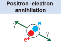
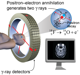
 The properties and physical location of radioactive nuclei can be observed by detecting the products of their decay with specific detectors. Many useful nuclear reactions release high-energy photons also called γ rays (gamma rays). Detecting these γ rays is a very well developed technology and it is used in many applications including medical diagnostic imaging. Radioactive nuclei such as (fluorine-18) decay by emitting a positron. (This process is a type of β decay in which a positron is emitted instead of an electron.) When a positron and an electron come into contact, which happens almost instantaneously in matter, they immediately “disappear” or annihilate each other. This annihilation reaction generates two γ rays, each with an energy of 0.511 MeV, which are very penetrating and can be detected with γ ray detectors.
|
 Engineers have designed special systems that detect these rays and map their origin in space. This technique is called positron emission tomography, or PET for short, and is used for high-resolution medical diagnostic imaging. The science of PET is based on the decay of , which has a half-life of 110 minutes. Since a half-life of 110 minutes is very short, does not occur naturally. Scientists have to produce these isotopes close to the place of use. To do so they use an accelerator to generate high-energy protons that interact with a target containing , which produces according to the reaction:
|
Once is produced it is ingested into the bloodstream. The isotopes concentrate in cells with high activity such as brain and tumor cells. Fluorine-18 then decays, releasing γ rays, which are detected by the γ-ray detectors surrounding the patient. The γ rays from the annihilation reaction are always produced in pairs that emit their energy in opposite directions. Special mathematical techniques and software have been developed that can construct an image of the region according to the concentration of . Radiologists examine the resulting images in the same way that they look at an x-ray image of an arm to see whether a bone is broken. By precisely mapping organs and tissues, a medical doctor can target treatment to the correct part of the body. 
|
Magnetic resonance imaging (MRI) takes advantage of a different nuclear property. The human body is full of water. Protons in the nucleus of hydrogen in the water molecule possess a quantum mechanical “spin” that will align itself with an outside magnetic field in a process called nuclear magnetic resonance. A magnetic resonance imaging machine has a powerful electromagnet that aligns the protons in the patient’s body in this way. The machine varies the electromagnetic field (using radio waves) to cause the protons to rotate around. As the protons rotate, they give off a unique signal that the MRI machine detects to visualize the patient’s internal tissues. 
|
| |
|

